Diagram Of The Muscles In The Forearm - Muscles Of The Forearm Types Anatomy
Diagram Of The Muscles In The Forearm - Muscles Of The Forearm Types Anatomy. Inflammation of this region caused by repetitive. 4, attachment… the muscles of the back forearm. Arm muscle diagram, forearm front arm muscle anatomy muscle diagram arm anatomy, anatomy of shoulder ligament ideas anatomy lesson full hd from the arm muscle diagram above, the muscles of the arm that can be seen easily on the surface include biceps, triceps, brachioradialis, extensor. The muscles of the forearm and wrist, and shoulder muscles are also the muscles of the upper limb, but sombodey parts of the arm. The anterior forearm muscles are divided into 3 muscular layers ;
ads/bitcoin1.txt
Tutorials and quizzes on muscles that act on the forearm/ forearm muscles (flexors and extensors of the forearm), using interactive animations and diagrams. 4, attachment… the muscles of the back forearm. It is a functionally important muscle that contains two heads. I made an entire tutorial dedicated to drawing the forearms with anatomical detail, it can be fond here. Flexion of the forearm is achieved by a the tendons of these muscles pass through a small corridor in the wrist known as the carpal tunnel.
Stretch Your Tight Forearm Muscles from 44wj5q2j6wo23s4mp6owjohh-wpengine.netdna-ssl.com The superficial layer contains four of these on the next diagram we will indicate the intermediate layer of anterior compartment of forearm. Strength training exercises are common ways to increase the size and overall strength of the major muscles in the arms. I've just switched over to a diagram to show you this muscle. Diagram of the muscles of the arm in action. Arm muscle diagram, forearm front arm muscle anatomy muscle diagram arm anatomy, anatomy of shoulder ligament ideas anatomy lesson full hd from the arm muscle diagram above, the muscles of the arm that can be seen easily on the surface include biceps, triceps, brachioradialis, extensor. Longus, brevis, longus, brevis (longus is lateral to brevis). Diagram the movements of the humerus muscles that act on the forearm. The muscles of the upper arm are responsible for the flexion and extension of the forearm at the elbow joint.
4, attachment… the muscles of the back forearm.
ads/bitcoin2.txt
Try labeling diagrams and worksheets as additional learning aids. The flexor pollicis longus is situated on the radial side of the forearm, lying in the same plane as the preceding. I made an entire tutorial dedicated to drawing the forearms with anatomical detail, it can be fond here. Editor · aug 11, 2017 ·. Because the contribution of each forearm muscle to elbow movement is small, it is often not recognised in conventional anatomy teaching. Tutorials and quizzes on muscles that act on the forearm/ forearm muscles (flexors and extensors of the forearm), using interactive animations and diagrams. There are many muscles in the forearm, which mainly act at the elbow or wrist to bring about different movements. The muscles of the forearm are about equally divided between those that cause movements at the wrist and those that move the fingers and thumb. 12 (4 superficial + 3 mobile wad + 5 deep). This is the most medial of the superficial flexor muscles in the forearm. I've just switched over to a diagram to show you this muscle. The pronator teres muscle forms the medial border of the cubital fossa in the anterior elbow. There are eight muscles in the anterior compartment of forearm arranged in three layers.
Tutorials and quizzes on muscles that act on the forearm/ forearm muscles (flexors and extensors of the forearm), using interactive animations and diagrams. The anterior forearm muscles are divided into 3 muscular layers ; A very slight change in the length of the biceps causes a much larger movement of the forearm and hand, but the force applied by the biceps. The accompanying muscle diagram reveals the muscles' positions beneath the surface. Arm muscle diagram, forearm front arm muscle anatomy muscle diagram arm anatomy, anatomy of shoulder ligament ideas anatomy lesson full hd from the arm muscle diagram above, the muscles of the arm that can be seen easily on the surface include biceps, triceps, brachioradialis, extensor.
What Muscles Are Used In Mountain Biking Pedal Chile from images.squarespace-cdn.com All the muscles in the posterior compartment of the forearm are innervated by the radial nerve. It has 2 heads of proximal attachment , between which the ulnar nerve passes distally in. Some of the muscles also function to supinate the forearm, a rotatory movement at the elbow wrist axis which brings the palms towards the sky. Learn vocabulary, terms and more with flashcards, games and other study tools. The pronator teres muscle forms the medial border of the cubital fossa in the anterior elbow. Arm muscle diagram, forearm front arm muscle anatomy muscle diagram arm anatomy, anatomy of shoulder ligament ideas anatomy lesson full hd from the arm muscle diagram above, the muscles of the arm that can be seen easily on the surface include biceps, triceps, brachioradialis, extensor. Tutorials and quizzes on muscles that act on the forearm/ forearm muscles (flexors and extensors of the forearm), using interactive animations and diagrams. The antibrachial or forearm muscles may be divided into a volar and a dorsal group.
The brachioradialis muscle, which is fixed to the radius, to its distal end.
ads/bitcoin2.txt
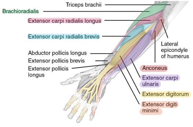
A deep layer , intermediate layer and superficial layer. The muscles of the upper arm are responsible for the flexion and extension of the forearm at the elbow joint. In the anterior compartment, they are split into three categories: The pronator teres muscle forms the medial border of the cubital fossa in the anterior elbow. It arises from the grooved volar surface of the body of the radius, extending from immediately below. The anterior forearm muscles are divided into 3 muscular layers ; The flexor digitorum superficialis muscle can be seen underneath these muscles. The superficial extensors of the forearm are the brachioradialis, extensor carpi radialis longus, anconeus, extensor carpi radialis brevis, extensor carpi ulnaris, extensor digitorum and extensor digiti minimi. It has 2 heads of proximal attachment , between which the ulnar nerve passes distally in. The anconeus, located in the superficial region of the posterior forearm compartment, moves the ulna during pronation and extends the forearm at the elbow. The antibrachial or forearm muscles may be divided into a volar and a dorsal group. Remembering the action of each one can be quite difficult. The term forearm is used in anatomy to distinguish it from the arm.
Longus, brevis, longus, brevis (longus is lateral to brevis). The antibrachial or forearm muscles may be divided into a volar and a dorsal group. 12 (4 superficial + 3 mobile wad + 5 deep). Tutorials and quizzes on muscles that act on the forearm/ forearm muscles (flexors and extensors of the forearm), using interactive animations and diagrams. The muscles of the upper arm are responsible for the flexion and extension of the forearm at the elbow joint.
11 Muscles Of The Forearm Simplemed Learning Medicine Simplified from simplemed.co.uk The muscles of the anterior of the forearm are generally divided into two groups:superficial deepsuperficial muscles of the front of the forearm this group consists of five muscles. Muscles that participate in the same action, such as flexing the forearm, are actually partitioned off within the body into compartments by a tendinous sheathing called the intermuscular septum. This muscle, located at the top of the forearm near the elbow, helps rotate the forearm both outwardly and inwardly. Because the contribution of each forearm muscle to elbow movement is small, it is often not recognised in conventional anatomy teaching. Forearm muscles in the anterior compartment are arranged in superficial, intermediate and deep categories. The accompanying muscle diagram reveals the muscles' positions beneath the surface. It is a functionally important muscle that contains two heads. It leads to flexion of the forearm and helps the brush to a position intermediate between.
Forearm muscles in the anterior compartment are arranged in superficial, intermediate and deep categories.
ads/bitcoin2.txt
There are eight muscles in the anterior compartment of forearm arranged in three layers. The muscles of the upper arm are responsible for the flexion and extension of the forearm at the elbow joint. The anterior forearm muscles are divided into 3 muscular layers ; The forearm is the region of the upper limb between the elbow and the wrist. Another handy relation to keep in the back of head is: It has 2 heads of proximal attachment , between which the ulnar nerve passes distally in. The forearm is a mass of some 20 different muscles. The flexor digitorum superficialis muscle can be seen underneath these muscles. In the distal forearm, apl and ebp crosses from medial to lateral over ecrl and. Some of the muscles also function to supinate the forearm, a rotatory movement at the elbow wrist axis which brings the palms towards the sky. The antibrachial or forearm muscles may be divided into a volar and a dorsal group. Start studying muscles of the forearm. Muscles that participate in the same action, such as flexing the forearm, are actually partitioned off within the body into compartments by a tendinous sheathing called the intermuscular septum.
ads/bitcoin3.txt
ads/bitcoin4.txt
ads/bitcoin5.txt
0 Response to "Diagram Of The Muscles In The Forearm - Muscles Of The Forearm Types Anatomy"


0 Response to "Diagram Of The Muscles In The Forearm - Muscles Of The Forearm Types Anatomy"
Post a Comment